

Short description and discussion of imaging findings:
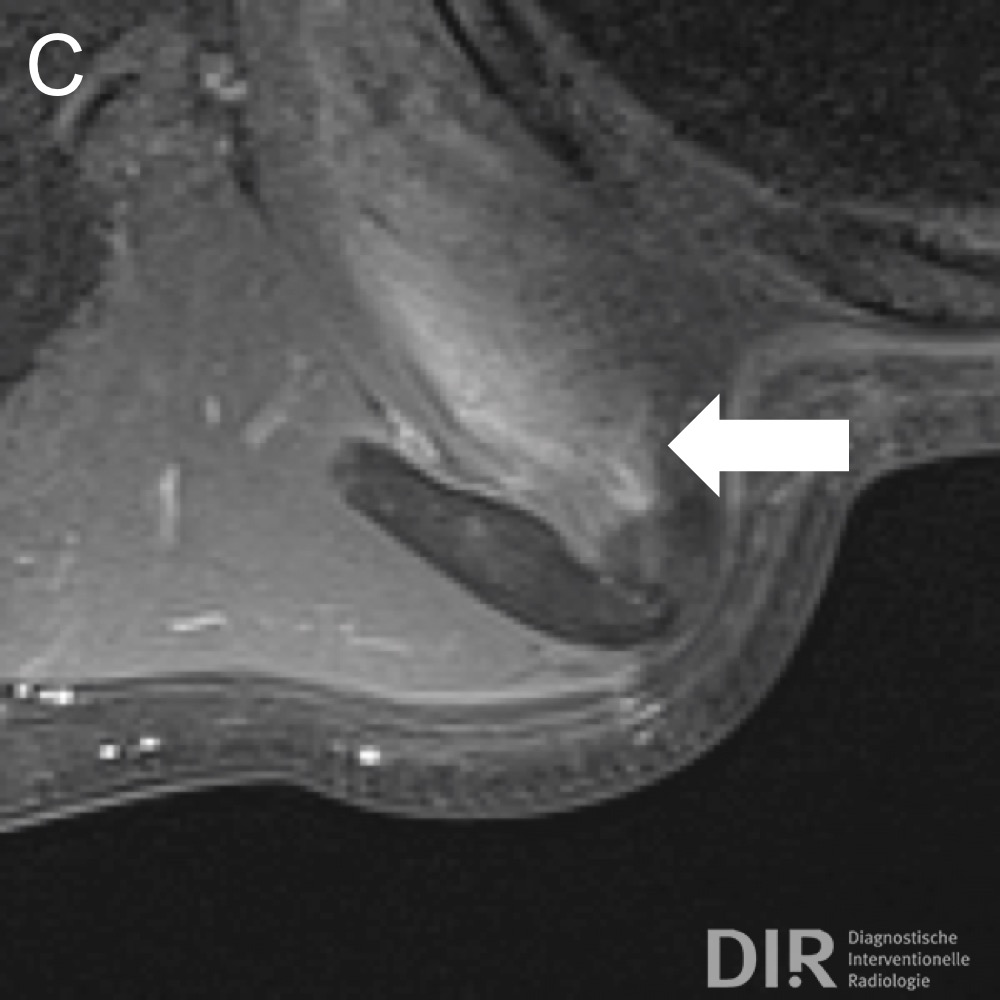
This 44 years old male presented with a slightly painful swelling of the right subscapular region. MRI of the right thoracic wall shows an ill-defined, lenticular, uncapsulated, heterogeneous mass slightly displacing the serratus anterior muscle compatible with elastofibroma dorsi. The lesion has hypointense to intermediate signal intensity in T1-weighting (A). Most of the mass has a signal intensity similar to that of skeletal muscle and fewer parts have some heterogeneous hyperintense signals in fat-suppressed T2 weighting (B). After contrast media application the lesion shows faint heterogeneous enhancement (C). Elastofibroma dorsi is a rare benign fibroelastic tumor-like condition made up of enlarged and irregular elastic fibers. Patients are usually elderly (mean age 65-70 years) with predominance of the female gender (5:1). In most cases it is unilateral, but in up to 10% it occurs bilaterally. The pathogenesis is not clear and theories about its origin include a genetic predisposition, repeated trauma and friction, and enzymatic defects resulting in abnormal elastogenesis (if occurring multifocal).
Certainty of diagnosis:
Pathognomic imaging findings
Recommended reading:
DOI: 10.1155/2008/756565


